Retinal adaptive optics imaging goes multimodal
4 July 2022
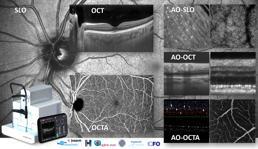
A publication in Nature Scientific Reports introduces a technology that delivers large-field views of the retina with several imaging modalities, as well as close-up views with 3-D cellular resolution.
The authors describe a prototype that integrates adaptive optics with scanning laser ophthalmoscopy, optical coherence tomography (OCT), and OCT angiography. The article also presents clinical images acquired in healthy volunteers and patients.
“In addition to multimodal retinal exams, this device allows us to see how lesions develop at the cellular scale. It is an extraordinary tool for understanding disease mechanisms and developing new therapies.”
Prof. Michael Larsen
Ophthalmologist at Copenhagen University Rigshospitalet
The new imaging system was developed by MERLIN, a collaborative project carried out by three academic teams, two clinical research centers and Imagine Eyes. This project has received financial support from the European Commission’s Horizon 2020 program.
“I am thrilled by the near-histological resolution of this new technology. Beyond revealing the effects of diseases and treatments on retinal cells, there is also a huge potential for precision assessments of retinal circulation.”
Prof. Michel Paques,
Ophthalmologist at Paris Quinze-Vingts Hospital
Article reference: Shirazi, M. F., Andilla, J., Lefaudeux, N. et al. (2022). Multi-modal and multi-scale clinical retinal imaging system with pupil and retinal tracking. Scientific Reports, 12(1), 9577
The MERLIN project has received € 4.868M in funding from the European Union’s Horizon 2020 research and innovation program, under grant agreement No 780989.
![]()

