Sensitive assessment of retinal structure and function in Usher syndrome
Published in Frontiers in Ophthalmology, a clinical study from University of Tuebingen investigates retinal function and structure at the cellular level in retinitis pigmentosa associated with Usher syndrome. It reports findings from rtx1 adaptive optics imaging (AO) and multifocal electroretinography (mfERG) in patients with various stages of RP.
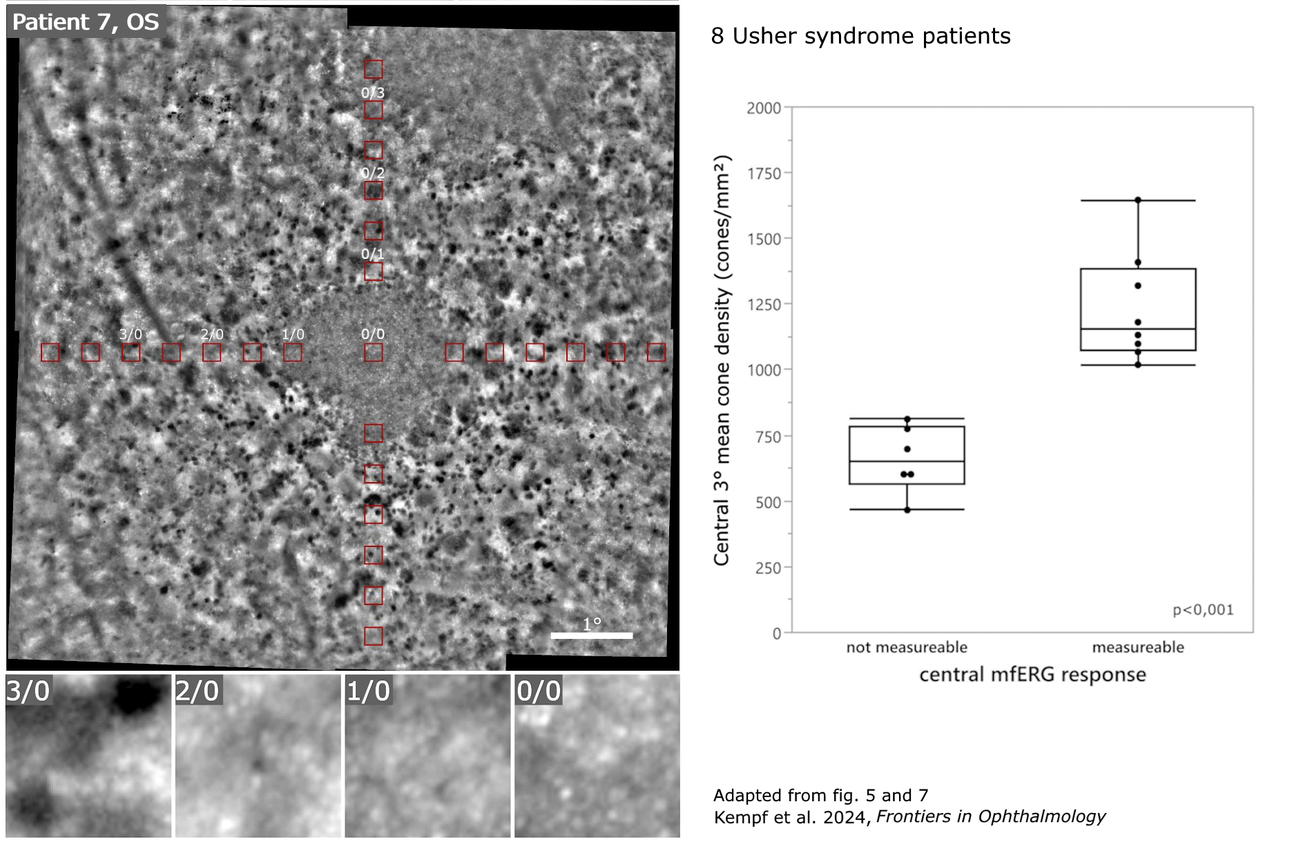
The rtx1 revealed different phenotypes of photoreceptor degeneration, with visible cells in the foveal area, allowing for quantitative analysis in the central 3 degrees.
“When the cone density measurements from AO retinal images were compared with the mfERG recordings from corresponding areas, the analysis indicated that there was a clear correlation between the strength of mfERG responses and the cone density measurements”
In some eyes with advanced RP, although mfERG was not recordable, the density of remaining cone cells could still be measured on rtx1 images.
Article reference: Kempf, M., Kohl, S., Stingl, K., Nasser, F., Stingl, K., & Kortuem, F. C. (2024). Adaptive optics retinal imaging in patients with usher syndrome. Fontiers in Ophthalmology, 4:1349234. doi: 10.3389/fopht.2024.1349234
