Vascular changes in glaucoma assessed with micron precision
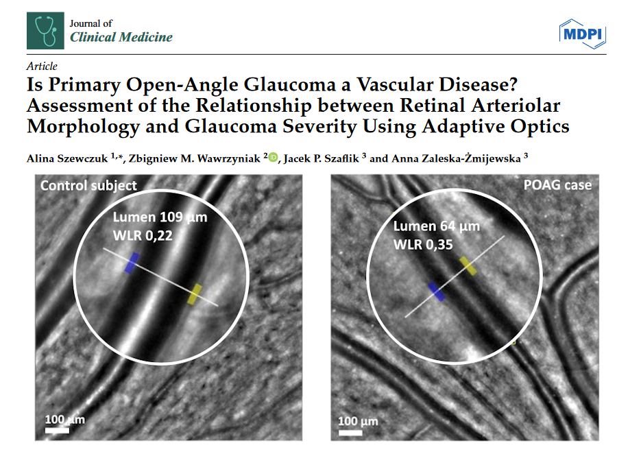
A new study published in Journal of Clinical Medicine reports alteration in retinal vessel structure in primary open angle glaucoma – even at early stages. Clinical researchers at Medical University of Warsaw used a rtx1 AO camera to capture the walls of retinal arterioles and extract micron-precision measurements.
“The high resolution of AO images makes them an ideal reference target for retinal vessel morphology measurements, with excellent intra-observer and inter-observer repeatability and a good correlation with SD-OCT measurements.”
Article reference: Szewczuk, A., Wawrzyniak, Z. M., Szaflik, J. P., & Zaleska-Żmijewska, A. (2024). Is Primary Open-Angle Glaucoma a Vascular Disease? Assessment of the Relationship between Retinal Arteriolar Morphology and Glaucoma Severity Using Adaptive Optics. Journal of Clinical Medicine, 13(478). https://doi.org/10.3390/jcm13020478