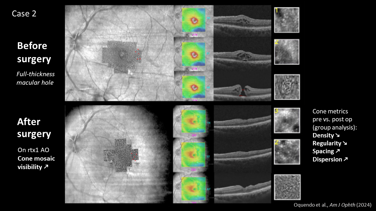
Cone cells before/after macular hole surgery
2024 has seen many new clinical insights and advances in therapies for retinal conditions.
We are pleased to share another study published in the American Journal of Ophthalmology.
To investigate the often incomplete visual recovery following macular hole surgery, researchers at the University of Toronto used a rtx1 AO retinal camera to measure visual cone cells before and after surgery.
“The high-resolution assessment of photoreceptors with AO imaging may be useful for future studies assessing the efficacy of surgical techniques on restoration of retinal anatomic integrity. Additionally, AO imaging has the potential to provide useful imaging biomarkers that predict postoperative functional outcomes.”

Article reference: Oquendo, P. L., Wright, T., Naidu, S. C., Pimentel, M. C., Hamli, H., Issa, M., Faleel, A., Nagel, F., Yan, P., & Muni, R. H. (2024). Comparison of the Photoreceptor Mosaic Before and After Macular Hole Surgery with High Resolution Adaptive Optics Imaging. American Journal of Ophthalmology, S0002-9394(24)00487-2. https://www.ajo.com/article/S0002-9394(24)00487-2/fulltext